We use cookies to improve your browsing experience and provide meaningful content. Read our cookie policy. Accept
  •  Customer Login
  • Register
  •  View Cart (0)
  •  Customer Login
  • Register
  •  View Cart (0)

Takara Bio
  • Products
  • Services & Support
  • Learning centers
  • APPLICATIONS
  • About
  • Contact Us

Clontech Takara Cellartis

Close

  • ‹ Back to Gene editing in hiPS cells
  • Tagging an endogenous gene with AcGFP1 in hiPS cells
  • Tagging an endogenous gene with a myc tag in hiPS cells
  • Generating clonal hiPS cell lines deficient in CD81
  • Introducing a tyrosinemia-related SNP in hiPS cells
  • Inserting an expression cassette into the AAVS1 locus in hiPS cells
  • Editing hiPS cells using electroporation
  • Editing hiPS cells using gesicle technology
  • Single-cell cloning of hiPS cells
DEF-CS culture system DEF-CS product page
Home › Learning centers › Stem cell research › Technical notes › Gene editing in hiPS cells › Editing hiPS cells using electroporation

Technical notes

  • Pluripotent stem cells
    • Using the DEF-CS system to culture human iPS cells
    • Comparison of the Cellartis DEF-CS system with other vendors' human iPS cell culture systems
    • Reprogramming PBMCs
    • Reprogramming fibroblasts
  • Gene editing in hiPS cells
    • Tagging an endogenous gene with AcGFP1 in hiPS cells
    • Tagging an endogenous gene with a myc tag in hiPS cells
    • Generating clonal hiPS cell lines deficient in CD81
    • Introducing a tyrosinemia-related SNP in hiPS cells
    • Inserting an expression cassette into the AAVS1 locus in hiPS cells
    • Editing hiPS cells using electroporation
    • Editing hiPS cells using gesicle technology
    • Single-cell cloning of hiPS cells
  • Organoids
    • Retinal organoid differentiation from iPSCs cultured in the Cellartis DEF-CS 500 Culture System
    • Liver organoid differentiation from iPSCs for prediction of drug-induced liver injury
    • Generation of embryonic organoids using NDiff 227 neural differentiation medium
  • Beta cells
    • Beta cells for disease modeling
  • Hepatocytes
    • hiPS-HEP cells for disease modeling
    • hiPS-HEP cells for drug metabolism studies
    • Power medium for long-term human primary hepatocyte culture
    • iPS cell to hepatocyte differentiation system
  • Cardiomyocytes
    • Making engineered heart tissue with cardiomyocytes
  • Neural stem cells
    • RHB-A neural stem cell medium
New products
Need help?
Contact Sales
DEF-CS culture system DEF-CS product page
Tech Note

Editing hiPS cells using electroporation

  • Highly efficient gene editing in hiPS cells
    Electroporate Cas9 protein and sgRNA, avoiding genomic integration and reducing off-target effects
  • Maintenance of pluripotency after editing, single-cell cloning, and expansion
    hiPS cells maintain high levels (>90%) of pluripotency markers Oct-4, TRA-1-60, and SSEA-4
  • Stable karyotype from start to finish
    hiPS cells maintain a normal and stable karyotype throughout editing, single-cell cloning, and expansion

Introduction Results Conclusions Methods References

Introduction  

CRISPR/Cas9 is a revolutionary tool that offers simplicity and flexibility for gene editing experiments. Stem cells are ideal candidates for editing, as they are highly renewable and expandable, and they can differentiate into multiple cell types. Human induced pluripotent stem (hiPS) cells offer the greatest utility due to their practically unlimited expansion and differentiation potential.

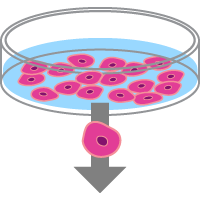
The sequential processes of somatic cell reprogramming to create patient-specific hiPS cells, CRISPR/Cas9 gene editing, and single-cell cloning uniquely enable researchers to study how a specific genetic modification can influence function. To create cell models for the discovery of disease etiology, progression, and treatment, isogenic cell lines can be created from healthy or sick individuals, then compared (Figure 1).     

Human iPS cells offer a renewable source of clonal diseased or healthy cells.  

Figure 1. Human iPS cells offer a renewable source of clonal diseased or healthy cells.

Cells from a healthy individual can be reprogrammed to create an expandable population of healthy hiPS cells. This healthy hiPS cell population can be edited to insert a known or theorized mutation and expanded clonally to yield complementary cell line(s) that only differ from the "healthy" cells by the introduced mutation. Later, this edited clonal cell line can be differentiated into a cell type relevant to the disease under study (e.g., neurons, as depicted in Figure 1). Alternatively, cells from a sick individual can be reprogrammed and expanded into a "diseased" hiPS cell line, then gene editing can be used to correct the mutation thought to cause the disease. These edited clonal cell lines can be differentiated into the cell type of interest as well. These renewable sources of diseased and healthy cell models, controlled for genetic variability, can be used for a variety of downstream applications to study and treat disease.

Challenges of editing hiPS cells and novel solutions

Despite the power and utility of CRISPR/Cas9 as an editing tool, some challenges must be overcome, particularly when editing hiPS cells. Delivery of Cas9 and the target-specific sgRNA requires an efficient method with low toxicity. Off-target effects should be minimized by the experimental design: choose a good sgRNA with the fewest possible off-target effects, control the amount of Cas9 introduced into the cells, and limit the time that Cas9 is present in the cells. Once edited, single cells with the desired mutation need to be expanded clonally, an inefficient process. Throughout all stages of editing and single-cell-expansion experiments, hiPS cells need to be highly proliferative and pluripotent.

To address the editing difficulties, we have developed a high-quality recombinant Cas9 protein, purified from E. coli, that is ready for use in electroporation experiments. Thus, there is no persistent expression of Cas9, because no coding gene is present. Combined with sgRNAs produced with our Guide-it In Vitro Transcription Kit, we consistently achieve high levels of functional gene knockouts and homology-directed repair (HDR) in hard-to-edit cells, including hematopoietic stem cells (HSCs) and hiPS cells.

To overcome the challenges with editing and single-cell cloning of hiPS cells, we have developed a method that combines the Cellartis iPSC Single-Cell Cloning DEF-CS Culture Media Kit with Guide-it Recombinant Cas9 (10 µg/µl). This method utilizes the DEF-CS culture system, recognized for its suitability for genome engineering (Valton et al. 2014) and single-cell cloning (Feng et al. 2014), to promote reliable growth of hiPS cells in a feeder-free and defined environment.

With this system, hiPS cells are grown as a homogeneous monolayer and are enzymatically passaged as single cells that maintain pluripotency with a stable karyotype for more than 20 passages (Asplund et al. 2016). The same reagents are used for monolayer and single-cell culture, reducing variability. When plated as single cells in wells of a 96-well plate, at least 50% of those seeded cells will proliferate into clonal colonies. In the following experiments, we demonstrate that the Cellartis iPSC Single-Cell Cloning DEF-CS Culture Media Kit combined with Guide-it Recombinant Cas9 (10 µg/µl) is ideal for performing footprint-free gene editing and single-cell cloning of edited hiPS cells.

Results  

Maintenance of pluripotency after editing: AcGFP1 knockout test case

An initial proof-of-concept experiment was performed to confirm that editing hiPS cells using electroporation does not influence pluripotency (Figure 2, Panel A). First, cells from Cellartis Human iPS Cell Line 22 (ChiPSC22) were modified to stably express AcGFP1, then these cells were cultured using the DEF-CS culture system in a 48-well plate. Next, we electroporated Guide-it Recombinant Cas9 in a ribonucleoprotein (RNP) complex with sgRNA specific to AcGFP1. The cells were then grown for nine days according to the Cellartis DEF-CS 500 Culture System User Manual. After, cells were analyzed by flow cytometry or immunocytochemistry for AcGFP1 and pluripotency markers. Untreated cells (negative control) were grown in parallel for the same amount of time as treated cells.

To determine pluripotency and AcGFP1 knockout efficiency in electroporated cells, cells were labeled with a fluorescently labeled antibody specific to the pluripotency markers Oct-4 and SSEA-4. Cells were analyzed via flow cytometry and the percentages of cells that were AcGFP1 positive Oct-4 positive, and SSEA-4 positive were quantified (Figure 2, Panel B). The negative control hiPS cells were ~94% AcGFP1 positive, ~96% Oct-4 positive, and over 99% SSEA-4 positive, indicating the cells were unedited and pluripotent. Treatment of hiPS cells by electroporating Cas9/sgRNA RNP complexes targeting AcGFP1 resulted in knockout of AcGFP1 expression in 90% of the cells. Furthermore, pluripotency-determined by Oct-4 and SSEA-4 expression-was maintained in 92% and 99% of analyzed cells, respectively.

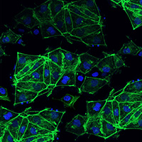
Additional confirmation of knockout and pluripotency was performed using immunocytochemistry (Figure 2, Panel C). Control- and electroporated hiPS cells were assessed for AcGFP1 expression (green), labeled with an antibody specific to Oct-4, visualized with a fluorescent-labeled secondary antibody (red), and nuclear-labeled with DAPI (blue). An isotype control for the secondary antibody was used as a negative control in both samples. In accordance with the flow cytometry data, expression of AcGFP1 in gesicle-treated cells was knocked out in the majority of cells, despite AcGFP1 being expressed in nearly all control cells. Nearly all hiPS cells were Oct-4 positive, supporting the conclusion that editing using electroporation does not alter pluripotency.

Electroporation knockout of AcGFP1 hiPS cells does not alter pluripotency when performed in the DEF-CS culture system.

Figure 2. Electroporation knockout of AcGFP1 hiPS cells does not alter pluripotency when performed in the DEF-CS culture system. Panel A. Electroporation delivery of Cas9/sgRNA ribonucleoproteins (RNPs) was used to knockout AcGFP1 in the Cellartis Human iPS Cell Line 22 stably expressing AcGFP1. Cells were cultured under non-differentiating conditions using the Cellartis DEF-CS 500 Culture System. After editing, cells were analyzed for AcGFP1 expression and pluripotency via flow cytometry (Panel B) and immunocytochemistry (Panel C). As a negative control, untreated parental cells were grown in parallel for the same amount of time. The quantification of AcGFP1 expression in the edited cells revealed a 90% knockout. Furthermore, pluripotency of the parental and edited cell populations was maintained, with nearly 100% of cells expressing SSEA-4 (assessed via flow cytometry in Panel B) and Oct-4 (assessed via flow cytometry in Panel B and immunohistochemistry in Panel C).  

Workflow for generating clonal hiPS cell lines deficient in CD81

After concluding from our proof-of-concept experiment that a successful electroporation-based knockout of a virally integrated gene can be achieved, we chose to target CD81, an endogenous membrane glycoprotein that forms complexes with integrins and plays a critical role in the infection process that leads to hepatitis C (Figure 3).

Workflow for targeted knockout of CD81

Figure 3. Workflow for targeted knockout of CD81, an endogenous gene in hiPS cells, using the Cellartis iPSC Single-Cell Cloning DEF-CS Culture Media Kit combined with Guide-it Recombinant Cas9 (10 µg/µl). hiPS cells can be cultured, edited, and clonally expanded using the Cellartis iPSC Single-Cell Cloning DEF-CS Culture Media Kit combined with Guide-it Recombinant Cas9 (10 µg/µl). Before and after using this system, hiPS cells are grown in the DEF-CS culture system, which maintains cells as a karyotypically stable and pluripotent monolayer.

Knockout of CD81 in cells from Cellartis Human iPS Cell Line 18 (ChiPSC18) was performed according to the Cellartis iPSC rCas9 Electroporation and Single-Cell Cloning System User Manual. Electroporation of Cas9 in an RNP complex with sgRNA specifically targeting CD81 was performed on the cells. Flow cytometry analysis of the total population was performed seven days later to determine the CD81 KO efficiency and overall pluripotency levels. Flow cytometry was employed to isolate the sorted population of CD81-negative cells. Single cells from this sorted population were then seeded into each well of a 96-well plate by limiting dilution. Single hiPS cells were expanded into clonal lines using the Cellartis iPSC Single-Cell Cloning DEF-CS Culture Media Kit and characterized for their pluripotency and karyotype.

Successful editing of human iPS cells and maintenance of pluripotency

After electroporation, assessment of CD81 expression by flow cytometry identified that 86.8% of electroporated hiPS cells were CD81 negative (Figure 4, Panel A). We also interrogated the population of sorted, CD81-negative cells for the expression of pluripotency markers (Figure 4, Panel B) and identified that successfully edited hiPS cells were 96.4% Oct-4 positive and 99.9% SSEA-4 positive. Taken together, these data indicate that hiPS cells expanded in the DEF-CS culture system can be successfully edited using electroporation while retaining their pluripotency.

CD81-negative hiPS cells remain pluripotent in the DEF-CS culture system. 

Figure 4. CD81-negative hiPS cells remain pluripotent in the DEF-CS culture system. Panel A. Electroporation of Cas9/sgRNA RNPs was used to knockout CD81 in the Cellartis Human iPS Cell Line 18. Cells were cultured under non-differentiating conditions using the Cellartis DEF-CS 500 Culture System. After editing, cells were analyzed for CD81 expression via flow cytometry. As a negative control, unedited parental cells were grown in parallel for the same amount of time. Quantification of CD81 expression in the electroporated cells revealed an 86.8% knockout efficiency. Panel B. CD81-negative cells were examined for pluripotency markers Oct-4 and SSEA-4. Pluripotency of parental (negative control) and edited (CD81–) cell populations was maintained, with 96.4% of edited cells expressing Oct-4 and ~100% of edited cells expressing SSEA-4.

Following electroporation, CD81-negative hiPS cells sorted via flow cytometry were counted using a hemocytometer. Cells were seeded as single cells using either limited dilution or flow cytometry. For flow cytometry, a single-cell was seeded into each well of a 96-well plate. Limiting dilution was performed by serial dilution until a final theoretical concentration of 50 cells/9,600 µl was achieved. Next, 100 µl of the final cell suspension was added to each well of a 96-well plate (reaching a final theoretical concentration of 0.5 cells/well). This increased the likelihood of obtaining wells containing only single cells and minimized the existence of doublets, which prevent the derivation of a clonal population. Following two weeks of culture, we identified between 31 and 33 emerging colonies from the single-cell seeding of ~50 cells using limited dilution, giving a calculated single-cell survival efficiency of ~62–66% (Table 1). hiPS cells seeded using flow cytometry had a slightly lower survival (52/96, 54%) likely due to the increased stress of passing through the flow cytometer into the plate. However, since seeding with the flow cytometer enabled more single cells to be plated, the overall number of emerging colonies was increased. These data demonstrate that the Cellartis iPSC rCas9 Electroporation and Single-Cell Cloning System enables highly efficient survival of single hiPS cells.

ChiPSC cell line

Isolation method Emerging colonies at 2 weeks from single cell Theoretical # of clones plated Calculated single-cell survival
ChiPSC 22 Limiting dilution 31 50 62%
ChiPSC 18 FACS 52 96 54%
ChiPSC 18 Limiting dilution 33 50 66%

Table I. Highly efficient survival of edited clones grown using the Cellartis iPSC Single-Cell Cloning DEF-CS Culture Media Kit. Typical rates for the generation of clonal colonies from a single-cell cloning experiment range from 1–5%. Using the Cellartis iPSC Single-Cell Cloning DEF-CS Culture Media Kit, we achieved an unparalleled survival rate of 54–66%.

Maintenance of pluripotency in expanded hiPS cell clones

Twelve of the emerging colonies from above were selected and expanded according to the user manual for the Cellartis iPSC rCas9 Electroporation and Single-Cell Cloning System. Once scaled up, each individual clonal line was assessed for pluripotency and knockout of CD81 using flow cytometry (Figure 5). All clones expressed high levels of Oct-4 (97–99% positive), TRA-1-60 (98–99% positive), and SSEA-4 (98–99% positive). Moreover, all lines were found to be CD81 deficient. These data show that pluripotency markers in the expanded clones are maintained at levels comparable to those in the parental line, ChiPSC18. Thus, using the Cellartis iPSC Single-Cell Cloning DEF-CS Culture Media Kit combined with Guide-it Recombinant Cas9 (10 µg/µl), we successfully knocked out endogenous CD81 from a starting line and generated 12 new, edited lines that were still pluripotent. 

Pluripotency was maintained in edited hiPS cell clones that were seeded as single cells.

Figure 5. Pluripotency was maintained in edited hiPS cell clones that were seeded as single cells. Individual, edited (CD81 knockout) hiPS cells were expanded into clonal lines and analyzed for expression of CD81 and three pluripotency markers via flow cytometry using antibodies against CD81, Oct-4, TRA-1-60, and SSEA-4. The parental hiPS cell line, ChiPSC18, was used as a control. As expected, all edited clones exhibited the loss of CD81 expression. Pluripotency was maintained in all edited clonal lines, as evidenced by the persistent expression of the three pluripotency markers.

Occurrence of a diverse set of indels in hiPS cell clones from the CD81 knockout experiment

We examined the specific base-pair (bp) insertions and deletions (indels) created during the CRISPR/Cas9 editing process in the clones using the Guide-it Indel Identification Kit. Because Cas9-induced double-strand breaks are mainly repaired via the error-prone, non-homologous-end-joining (NHEJ) DNA repair pathway, every cell that is edited should have a unique set of indels at the targeted gene-in this experiment, CD81. Accordingly, we saw a wide range of indels in the different clonal cell lines (Figure 6 and Table II). In some cases, CD81 knockout was accomplished via relatively small indels. For example, clone #1 had only a 1-bp insertion on both alleles at the editing site, and clone #10 had only a 1-bp deletion on both alleles at the editing site. Conversely, some indels were much larger. For example, clone #9 had an 53-bp insertion on one allele and a 51-bp insertion on the other allele. Taken together, these data demonstrate the diversity of indels created by CRISPR/Cas9 editing and highlight the utility in creating and screening multiple clones to account for this variability.  

Representation of indels created at the CD81 target site in different CRISPR/Cas9-edited hiPS cell clones.

Figure 6. Representation of indels created at the CD81 target site in different CRISPR/Cas9-edited hiPS cell clones. The genomic CD81 region of each clone was PCR amplified and sequenced using the Guide-it Indel Identification Kit to obtain detailed sequence information. The results show that each clone has a unique set of biallelic indels that knockout CD81. See Table II for a tabular representation of these data.

Diversity of indels in CRISPR/Cas9 gesicle-treated hiPS cell clones
Clone # 1 2 3 4 5 6 7 8 9 10 11 12
Indels (bp) Allele 1 +1 +3/
–26
–1 –1 –17 –21 –4 –8 +53 –1 –1 +1/–3
Allele 2 +1 –7 –7 +1 +1 –1 –18 –1 +51 –1 –11 –2

Table II. Tabular representation of the sizes and locations of indels created at the target site in CD81 for different CRISPR/Cas9-edited hiPS cell clones. Examples from this dataset are also shown in Figure 6. The results show that each clone has a unique set of indels that knockout CD81. For each allele, "+" values indicate the number of inserted nucleotides, while "–" values indicate the number of deleted nucleotides. If an insertion and deletion happened simultaneously, it is marked with a slash. If the insertion sequence corresponds to another part of the genome, that chromosome is shown between brackets. 

Normal karyotypes observed in edited hiPS cells

CRISPR/Cas9-mediated editing and single-cell cloning can be harsh on hiPS cells. Traditionally, these processes can force selective pressures that favor unintended mutations to the karyotype, conferring competitive advantages to in vitro growth. These karyotypic abnormalities render the cells unsuitable for study. Consequently, it is essential that the karyotype remains unaltered for multiple passages after editing.

To confirm karyotype stability, we examined four of the clonal lines, each of which had been expanded from one single cell to a confluent line in a 10-cm dish over a period of approximately one month (Figure 7). All lines were found to have normal, stable karyotypes. Thus, these data show that the Cellartis iPSC Single-Cell Cloning DEF-CS Culture Media Kit combined with Guide-it Recombinant Cas9 (10 µg/µl) effectively edits and expands single-cell hiPS cell clones without introducing karyotypic abnormalities.

hiPS cell clones that have been edited via electroporation maintain a stable karyotype after editing and clonal expansion

Figure 7. hiPS cell clones that have been edited via electroporation maintain a stable karyotype after editing and clonal expansion. The karyotypes of four edited clonal cell lines were analyzed. All clonal lines showed the expected 46, XY karyotype of the original ChiPSC18 cell line. Clonal cell line #6 is shown as an example.

Conclusions  

Combining the Cellartis iPSC Single-Cell Cloning DEF-CS Culture Media Kit with Guide-It Recombinant Cas9 (10 μg/μl) provides an efficient and effective method to generate clonal lines of edited hiPS cells. The system can enable high editing efficiency, with no discernible effect on hiPS cell health or pluripotency. Single-cell seeding of edited hiPS cells in this system results in high survival of pluripotent, edited clones, yielding a diverse set of edited clonal lines. Critically, the expanded lines maintain a normal karyotype, rendering the cells suitable for further investigation and use in screening and disease modeling.

Methods  

Cell culture

Cellartis human iPS cells from the ChiPSC22 line (Figure 2) and the ChiPSC18 line (Figures 3–7 and Table I) were grown in the Cellartis DEF-CS 500 Culture System (Cat. # Y30010) before editing. After single-cell cloning and expansion into 48-well plates with the Cellartis iPSC Single-Cell Cloning DEF-CS Culture Media Kit, clonal colonies were returned to the Cellartis DEF-CS 500 Culture System for further expansion prior to indel analysis (Figure 6 and Table II) and karyotyping (Figure 7). Please refer to the Cellartis DEF-CS 500 Culture System User Manual and the Cellartis iPSC rCas9 Electroporation and Single-Cell Cloning System User Manual for specific culture conditions and protocols.

Preparation of RNP complexes and electroporation

sgRNAs targeting AcGFP1 or CD81 were generated using the Guide-it sgRNA In Vitro Transcription Kit (Cat. # 632635) and purified using the Guide-it IVT RNA Clean-Up Kit (Cat. # 632638; also included as a part of Cat. # 632635). Guide-it Recombinant Cas9 (10 µg/µl) (Cat. # 632679) and purified sgRNAs were incubated to form RNP complexes and electroporated into ChiPSC18 cells using a Neon Transfection System (ThermoFisher Scientific). Please refer to the Guide-it Recombinant Cas9 (10 µg/µl) User Manual for specific protocols.  

Flow cytometry

Labeling of Oct-4 was performed by cell fixation and permeabilization followed by an incubation with anti-Oct-4-PE antibody (BD Pharmingen, Cat. #560186; 20 µl for 1 x 10⁶ cells) in PBS for 30 minutes. Cell labeling of extracellular proteins or markers was performed following standard labeling procedures. In Figure 4, Panel A, cells were incubated for 30 minutes with anti-SSEA-4-PE (BD Pharmingen, Cat. #560128; 20 µl for 1 x 10⁶ cells), anti-TRA-1-60-FITC (BD Pharmingen, Cat. #560380; 20 µl for 1 x 10⁶ cells), or anti-CD81-FITC (BD Pharmingen, Cat. #551108; 20 µl for 1 x 10⁶ cells) antibodies. In Figure 4, Panel B, cells were incubated simultaneously with anti-SSEA-4 and anti-TRA-1-60 antibodies, since they were labeled with different fluorophores (PE and FITC, respectively).

Immunocytochemistry

ChiPSC22 cells were grown in chamber slides until fixation with 4% paraformaldehyde. Cell permeabilization was achieved with 0.5% Triton X-100 for 5 minutes. After washing with PBS and blocking for 30 minutes (IHC/ICC Blocking Buffer - Low Protein; eBioscience, Cat. #00-4953), cells were incubated with anti-Oct-4 antibody (diluted 1:150; eBioscience , Cat. #41-5841) or an IgG2a K isotype control (diluted 1:150; eBioscience , Cat. #41-4321) for one hour. After the incubation period, cells were washed and mounted with anti-fading reagent containing DAPI (VECTASHIELD Antifade Mounting Medium with DAPI; Vector Laboratories, Cat. #H-1200).

Indel analysis

Indels within each edited clonal cell line were determined using the Guide-it Indel Identification Kit (Cat. #631444), following the kit's user manual.

Karyotyping

Analysis of the karyotypes of ChiPSC18 cell lines was performed by Cell Guidance Systems.

References  

Asplund, A. et al. One Standardized Differentiation Procedure Robustly Generates Homogenous Hepatocyte Cultures Displaying Metabolic Diversity from a Large Panel of Human Pluripotent Stem Cells. Stem Cell Rev. Reports 12, 90–104 (2016).   

Feng, Q. et al. Scalable Generation of Universal Platelets from Human Induced Pluripotent Stem Cells. Stem Cell Reports 3, 817–831 (2014).           

Valton, J. et al. Efficient strategies for TALEN-mediated genome editing in mammalian cell lines. Methods 69, 151–170 (2014).   

Related Products

Cat. # Product Size Price License Quantity Details
632679 Guide-it™ Recombinant Cas9 (10 µg/µl) 500 ug USD $847.00

License Statement

ID Number  
272 This product (“Product”) and its use, is the subject of U.S. Patents 8,697,359 and 8,771,945 and pending U.S. Patent applications. The purchase of the Product conveys to the buyer the non-transferable right to use Product(s) purchased from Takara Bio USA, Inc. or its Affiliates, and any progeny, modification or derivative of a Product, or any cell or animal made or modified through use of a Product, or any progeny, modification or derivative of such cell or animal (“Related Material”), solely for research conducted by the buyer in accordance with all of the following requirements. No right is given to use this Product or Related Material for any other purpose, including, but not limited to, use in drugs, in vitro diagnostic purposes, therapeutics, or in humans. (i) The buyer shall not sell or otherwise transfer Products (including without limitation any material that contains a Licensed Product in whole or part) or any Related Material to any other person or entity, or use Products or any Related Material to perform services for the benefit of any other person or entity, (ii) the buyer shall use only the purchased amount of the Products and components of the Products, and shall use any Related Material, only for its internal research and not for (a) the practice, performance or provision of any method, process or service, or (b) the manufacture, sale, use, distribution, disposition or importing of any product, in each case (a) or (b) for consideration, or on any other commercial basis (“Commercial Purpose”), (iii) the buyer shall use Licensed Products and any Related Material in compliance with all applicable laws and regulations, including without limitation applicable human health and animal welfare laws and regulations, and (iv) the buyer shall indemnify, defend and hold harmless MIT, Harvard and The Broad and their current and former trustees, directors, officers, faculty, affiliated investigators, students, employees, and agents and their respective successors, heirs and assigns (“Indemnitees”), against any liability, damage, loss, or expense (including without limitation reasonable attorneys’ fees and expenses) incurred by or imposed upon any of the Indemnitees in connection with any claims, suits, investigations, actions, demands or judgments arising out of or related to the exercise of any rights granted to the buyer, or any breach of the rights granted hereunder by the buyer.
391 LIMITED USE LABEL LICENSE: RESEARCH USE ONLY Notice to Purchaser: This product is the subject to a license granted to Takara Bio USA, Inc. and its Affiliates from Caribou Biosciences, Inc., and this product is transferred to the end-user purchaser (“Purchaser”) subject to a “Limited Use Label License” conveying to the Purchaser a limited, non-transferable right to use the product, solely as provided to Purchaser, together with (i) progeny or derivatives of the product generated by the Purchaser (including but not limited to cells), and (ii) biological material extracted or derived from the product or its corresponding progeny or derivatives (including but not limited to cells) (collectively, the product, and (i) and (ii) are referred to as (“Material”) only to perform internal research for the sole benefit of the Purchaser. The Purchaser cannot sell or otherwise transfer Material to a third party or otherwise use the Material for any Excluded Use. “Excluded Use” means any and all: (a) commercial activity including, but not limited to, any use in manufacturing (including but not limited to cell line development for purposes of bioproduction), product testing, or quality control; (b) preclinical or clinical testing or other activity directed toward the submission of data to the U.S. Food and Drug Administration, or any other regulatory agency in any country or jurisdiction where the active agent in such studies comprises the Material; (c) use to provide a service, information, or data to a third party; (d) use for human or animal therapeutic, diagnostic, or prophylactic purposes or as a product for therapeutics, diagnostics, or prophylaxis; (e) activity in an agricultural field trial or any activity directed toward the submission of data to the U.S. Department of Agriculture or any other agriculture regulatory agency; (f) high throughput screening drug discovery purposes (i.e., the screening of more than 10,000 experiments per day) as well as scale-up production activities for commercialization; (g) modification of human germline, including editing of human embryo genomes (with the sole exception of editing human embryonic stem (ES) cell lines for research purposes) or reproductive cells; (h) self-editing; and/or (i) stimulation of biased inheritance of a particular gene or trait or set of genes or traits (“gene drive”). It is the Purchaser’s responsibility to use the Material in accordance with all applicable laws and regulations. For information on obtaining additional rights, including commercial rights, please contact licensing@cariboubio.com or Caribou Biosciences, Inc., 2929 7th Street, Suite 105, Berkeley, CA 94710 USA, Attn: Licensing.
396 Sigma-Aldrich CRISPR Use License Agreement This Product and its use are the subject of one or more of the following issued patents and patent applications: Australia Patent Nos. 2013355214; 2017204031; and 2018229489; Canada Patent Nos. 2,891,347 and 2,977,152; China Patent No. CN105142669; European Patent Nos. EP 2 928 496 B1; EP 3 138 910 B1, 3 138 911 B1, EP 3 138 912 B1, EP 3 360 964 B1, EP 3 363 902 B1; Israel Patent No. IL238856; Singapore Patent No. 11201503824S; South Korea Patent Nos. 10-1844123 and 10-2006880; and U.S. Patent Application Serial Nos. 15/188,911; 15/188,924; 15/188,927; 15/188,931; and 15/456,204 (the “Patent Rights”). The purchase of this Product conveys to you (the “Buyer”) the NON-TRANSFERABLE right to use the Product for Licensed Research Use (see definition below) subject to the conditions set out in this License Agreement. 1. “Licensed Research Use” means any use for research purposes, except: (i) Buyer may not sell or otherwise transfer the Product (including without limitation any material that contains the Product in whole or part) or any Related Material to any other third party (except that you may transfer the Product, or any Related Material to a bona fide collaborator or contract research organization), or use the Products or any Related Material to perform services for the benefit of any other third party; (ii) Buyer may use only the purchased amount of the Product and components of the Product, and shall use any Related Material, only for your internal research within the Field, and not for any Commercial Purposes; (iii) Buyer shall use the Product and any Related Material in compliance with all applicable laws and regulations, including without limitation applicable human health and animal welfare laws and regulations; and (iv) the Buyer shall indemnify, defend, and hold harmless SIGMA and their current and former directors, officers, employees and agents, and their respective successors, heirs and assigns (the “Indemnities”) against any liability, damage, loss, or expense (including without limitation reasonable attorneys’ fees and expenses) incurred by or imposed upon any of the Indemnitees in connection with any claims, suits, investigations, actions, demands or judgments arising out of or related to the exercise of any rights granted to the Buyer hereunder or any breach of this License Agreement by such Buyer. 2. For purposes of Section 1 above, the following definitions shall apply: “Commercial Purposes” means (a) the practice, performance or provision of any method, process or service, or (b) the manufacture, sale, use, distribution, disposition or importing of any product, in each case (a) or (b) for consideration, or on any other commercial basis. “Field” means use as a research tool for research purposes; provided, however, that notwithstanding the foregoing, the Field shall expressly exclude (a) any in vivo and ex vivo human or clinical use, including, without limitation, any administration into humans or any diagnostic or prognostic use, (b) the creation of transgenic rodent models and/or derivatives thereof (including, but not limited to, rodents’ cells and rodents’ organs) by for-profit entities, (c) any in vivo veterinary or livestock use, or non-research agricultural use, or (d) use as a testing service, therapeutic or diagnostic for humans or animals. “Related Materials” means any progeny, modification or derivative of a Product. 3. Your right to use the Product will terminate immediately if you fail to comply with these terms and conditions. You shall, upon such termination of your rights, destroy all Product, Related Materials, and components thereof in your control, and notify SIGMA of such in writing. For information on purchasing a license to this Product for purposes other than Licensed Research Use, contact your local SIGMA sales representative, or call +1 800-325-3010.

Guide-it Recombinant Cas9 (10 µg/µl) is a recombinant wild-type Streptococcus pyogenes Cas9 nuclease expressed with a C-terminal nuclear localization signal (NLS), provided in 50% glycerol. The Cas9 protein solution has been verified to be sterile and well-tolerated by mammalian cells when electroporated as a ribonucleoprotein complex (RNP) with a single guide RNA (sgRNA) for knockout experiments, or as an RNP with a donor repair template for knockin experiments.

Notice to purchaser

Our products are to be used for Research Use Only. They may not be used for any other purpose, including, but not limited to, use in humans, therapeutic or diagnostic use, or commercial use of any kind. Our products may not be transferred to third parties, resold, modified for resale, or used to manufacture commercial products or to provide a service to third parties without our prior written approval.

Documents Components Image Data

Back

Knock out of CD81 in induced pluripotent stem cells

Knock out of CD81 in induced pluripotent stem cells
Knock out of CD81 in induced pluripotent stem cells. In this experiment, two induced pluripotent stem cell lines, hiPSC-18 and hiPSC-22, were electroporated withCas9/sgRNA RNPs against CD81. Cells were stained for CD81 and then run on a FACS machine to detect CD81 ten days after editing. The negative staining control shows the FACS data from cells without antibody staining, while the electroporation control shows cells electroporated without Cas9/sgRNA RNPs. When cells were electroporated with both Cas9 and sgRNA, very high editing efficiency was observed.

.

Back

Homology-directed repair at the AAVS1 or CXCR4 genes

Homology-directed repair at the AAVS1 or CXCR4 genes

Homology-directed repair at the AAVS1 or CXCR4 genes. Panel A. These diagrams demonstrate how the HDR experiments in CD34+ HSCs were done. A ssdNA template containing a HindIII restriction site was inserted into the AAVS1 gene, and a template containing both HindIII and BamHI restriction sites was inserted into the CXCR4 gene. The homology arms were 90 bp on each side. Panel B. After electroporation of the repair template and Cas9/sgRNA RNPs, gene targets were amplified from target cells using PCR and analyzed by restriction digest. Editing efficiencies are shown above each positive well on the gel. The extra band in the BamHI digest of the CXCR4 gene is due to a second BamHI site already present in the wild-type gene before editing. We verified that 98% of the HSCs stained positive for CD34+ five days after editing.

.

Back

Gene knockouts in the Cellartis hiPSC-18 cell line

Gene knockouts in the Cellartis hiPSC-18 cell line

Gene knockouts in the Cellartis hiPSC-18 cell line. The strength of the cleaved bands gives a semiquantitative estimate of the percentage of edited cells. As can be seen, the Guide-it recombinant Cas9, when combined with sgRNA from the Guide-it In Vitro Transcription Kit, provides consistent and effective gene editing for many targets when compared with other vendors’ recommended methods for producing guide RNAs. Numbers at the bottom of each gel represent gene editing efficiencies (expressed as a %) for the indicated RNPs.

Back

Knockout of therapeutically relevant genes in CD3+ Pan T cells

Knockout of therapeutically relevant genes in CD3+ Pan T cells

Knockout of therapeutically relevant genes in CD3+ Pan T cells. In this experiment, CD3+ Pan T cells were electroporated with Cas9/sgRNA RNPs in order to perform single (B2M or PD-1) or double knockouts (B2M+PD-1 or TRAC+TRBC). After thawing, cells were grown in LymphoONE medium and activated with ImmunoCult Human CD3/CD28/CD2 T Cell Activator for three days. Cells were then electroporated with RNP complexes in triplicate, following the Guide-it Recombinant Cas9 (10 μg/μl) User Manual. For the double knockouts, RNP complexes targeting the respective genes were prepared independently and mixed with the cells just before electroporation. Seven days after electroporation, knockout frequencies were measured by flow cytometry.

Back

Knockout of B2M in CD3+ Pan T cells

Knockout of B2M in CD3+ Pan T cells

Knockout of B2M in CD3+ Pan T cells. In this experiment, CD3+ Pan T cells were electroporated with Cas9/sgRNA RNPs in order to knockout B2M. After thawing, cells were grown in LymphoONE medium and activated with ImmunoCult Human CD3/CD28/CD2 T Cell Activator for three days. Cells were then electroporated with RNP complexes in triplicate, according to the Guide-it Recombinant Cas9 (10 μg/μl) User Manual. Seven days after electroporation, knockout frequencies and cell viability were measured by flow cytometry.

Back

632679: Guide-it Recombinant Cas9 (10 µg/µl)

632679: Guide-it Recombinant Cas9 (10 µg/µl)
632678 Guide-it™ Recombinant Cas9 (10 µg/µl) 200 ug USD $458.00

License Statement

ID Number  
272 This product (“Product”) and its use, is the subject of U.S. Patents 8,697,359 and 8,771,945 and pending U.S. Patent applications. The purchase of the Product conveys to the buyer the non-transferable right to use Product(s) purchased from Takara Bio USA, Inc. or its Affiliates, and any progeny, modification or derivative of a Product, or any cell or animal made or modified through use of a Product, or any progeny, modification or derivative of such cell or animal (“Related Material”), solely for research conducted by the buyer in accordance with all of the following requirements. No right is given to use this Product or Related Material for any other purpose, including, but not limited to, use in drugs, in vitro diagnostic purposes, therapeutics, or in humans. (i) The buyer shall not sell or otherwise transfer Products (including without limitation any material that contains a Licensed Product in whole or part) or any Related Material to any other person or entity, or use Products or any Related Material to perform services for the benefit of any other person or entity, (ii) the buyer shall use only the purchased amount of the Products and components of the Products, and shall use any Related Material, only for its internal research and not for (a) the practice, performance or provision of any method, process or service, or (b) the manufacture, sale, use, distribution, disposition or importing of any product, in each case (a) or (b) for consideration, or on any other commercial basis (“Commercial Purpose”), (iii) the buyer shall use Licensed Products and any Related Material in compliance with all applicable laws and regulations, including without limitation applicable human health and animal welfare laws and regulations, and (iv) the buyer shall indemnify, defend and hold harmless MIT, Harvard and The Broad and their current and former trustees, directors, officers, faculty, affiliated investigators, students, employees, and agents and their respective successors, heirs and assigns (“Indemnitees”), against any liability, damage, loss, or expense (including without limitation reasonable attorneys’ fees and expenses) incurred by or imposed upon any of the Indemnitees in connection with any claims, suits, investigations, actions, demands or judgments arising out of or related to the exercise of any rights granted to the buyer, or any breach of the rights granted hereunder by the buyer.
391 LIMITED USE LABEL LICENSE: RESEARCH USE ONLY Notice to Purchaser: This product is the subject to a license granted to Takara Bio USA, Inc. and its Affiliates from Caribou Biosciences, Inc., and this product is transferred to the end-user purchaser (“Purchaser”) subject to a “Limited Use Label License” conveying to the Purchaser a limited, non-transferable right to use the product, solely as provided to Purchaser, together with (i) progeny or derivatives of the product generated by the Purchaser (including but not limited to cells), and (ii) biological material extracted or derived from the product or its corresponding progeny or derivatives (including but not limited to cells) (collectively, the product, and (i) and (ii) are referred to as (“Material”) only to perform internal research for the sole benefit of the Purchaser. The Purchaser cannot sell or otherwise transfer Material to a third party or otherwise use the Material for any Excluded Use. “Excluded Use” means any and all: (a) commercial activity including, but not limited to, any use in manufacturing (including but not limited to cell line development for purposes of bioproduction), product testing, or quality control; (b) preclinical or clinical testing or other activity directed toward the submission of data to the U.S. Food and Drug Administration, or any other regulatory agency in any country or jurisdiction where the active agent in such studies comprises the Material; (c) use to provide a service, information, or data to a third party; (d) use for human or animal therapeutic, diagnostic, or prophylactic purposes or as a product for therapeutics, diagnostics, or prophylaxis; (e) activity in an agricultural field trial or any activity directed toward the submission of data to the U.S. Department of Agriculture or any other agriculture regulatory agency; (f) high throughput screening drug discovery purposes (i.e., the screening of more than 10,000 experiments per day) as well as scale-up production activities for commercialization; (g) modification of human germline, including editing of human embryo genomes (with the sole exception of editing human embryonic stem (ES) cell lines for research purposes) or reproductive cells; (h) self-editing; and/or (i) stimulation of biased inheritance of a particular gene or trait or set of genes or traits (“gene drive”). It is the Purchaser’s responsibility to use the Material in accordance with all applicable laws and regulations. For information on obtaining additional rights, including commercial rights, please contact licensing@cariboubio.com or Caribou Biosciences, Inc., 2929 7th Street, Suite 105, Berkeley, CA 94710 USA, Attn: Licensing.
396 Sigma-Aldrich CRISPR Use License Agreement This Product and its use are the subject of one or more of the following issued patents and patent applications: Australia Patent Nos. 2013355214; 2017204031; and 2018229489; Canada Patent Nos. 2,891,347 and 2,977,152; China Patent No. CN105142669; European Patent Nos. EP 2 928 496 B1; EP 3 138 910 B1, 3 138 911 B1, EP 3 138 912 B1, EP 3 360 964 B1, EP 3 363 902 B1; Israel Patent No. IL238856; Singapore Patent No. 11201503824S; South Korea Patent Nos. 10-1844123 and 10-2006880; and U.S. Patent Application Serial Nos. 15/188,911; 15/188,924; 15/188,927; 15/188,931; and 15/456,204 (the “Patent Rights”). The purchase of this Product conveys to you (the “Buyer”) the NON-TRANSFERABLE right to use the Product for Licensed Research Use (see definition below) subject to the conditions set out in this License Agreement. 1. “Licensed Research Use” means any use for research purposes, except: (i) Buyer may not sell or otherwise transfer the Product (including without limitation any material that contains the Product in whole or part) or any Related Material to any other third party (except that you may transfer the Product, or any Related Material to a bona fide collaborator or contract research organization), or use the Products or any Related Material to perform services for the benefit of any other third party; (ii) Buyer may use only the purchased amount of the Product and components of the Product, and shall use any Related Material, only for your internal research within the Field, and not for any Commercial Purposes; (iii) Buyer shall use the Product and any Related Material in compliance with all applicable laws and regulations, including without limitation applicable human health and animal welfare laws and regulations; and (iv) the Buyer shall indemnify, defend, and hold harmless SIGMA and their current and former directors, officers, employees and agents, and their respective successors, heirs and assigns (the “Indemnities”) against any liability, damage, loss, or expense (including without limitation reasonable attorneys’ fees and expenses) incurred by or imposed upon any of the Indemnitees in connection with any claims, suits, investigations, actions, demands or judgments arising out of or related to the exercise of any rights granted to the Buyer hereunder or any breach of this License Agreement by such Buyer. 2. For purposes of Section 1 above, the following definitions shall apply: “Commercial Purposes” means (a) the practice, performance or provision of any method, process or service, or (b) the manufacture, sale, use, distribution, disposition or importing of any product, in each case (a) or (b) for consideration, or on any other commercial basis. “Field” means use as a research tool for research purposes; provided, however, that notwithstanding the foregoing, the Field shall expressly exclude (a) any in vivo and ex vivo human or clinical use, including, without limitation, any administration into humans or any diagnostic or prognostic use, (b) the creation of transgenic rodent models and/or derivatives thereof (including, but not limited to, rodents’ cells and rodents’ organs) by for-profit entities, (c) any in vivo veterinary or livestock use, or non-research agricultural use, or (d) use as a testing service, therapeutic or diagnostic for humans or animals. “Related Materials” means any progeny, modification or derivative of a Product. 3. Your right to use the Product will terminate immediately if you fail to comply with these terms and conditions. You shall, upon such termination of your rights, destroy all Product, Related Materials, and components thereof in your control, and notify SIGMA of such in writing. For information on purchasing a license to this Product for purposes other than Licensed Research Use, contact your local SIGMA sales representative, or call +1 800-325-3010.

Guide-it Recombinant Cas9 (10 µg/µl) is a recombinant wild-type Streptococcus pyogenes Cas9 nuclease expressed with a C-terminal nuclear localization signal (NLS), provided in 50% glycerol. The Cas9 protein solution has been verified to be sterile and well-tolerated by mammalian cells when electroporated as a ribonucleoprotein complex (RNP) with a single guide RNA (sgRNA) for knockout experiments, or as an RNP with a donor repair template for knockin experiments.

Notice to purchaser

Our products are to be used for Research Use Only. They may not be used for any other purpose, including, but not limited to, use in humans, therapeutic or diagnostic use, or commercial use of any kind. Our products may not be transferred to third parties, resold, modified for resale, or used to manufacture commercial products or to provide a service to third parties without our prior written approval.

Documents Components Image Data

Back

632678: Guide-it Recombinant Cas9 (10 µg/µl)

632678: Guide-it Recombinant Cas9 (10 µg/µl)

Back

Knock out of CD81 in induced pluripotent stem cells

Knock out of CD81 in induced pluripotent stem cells
Knock out of CD81 in induced pluripotent stem cells. In this experiment, two induced pluripotent stem cell lines, hiPSC-18 and hiPSC-22, were electroporated withCas9/sgRNA RNPs against CD81. Cells were stained for CD81 and then run on a FACS machine to detect CD81 ten days after editing. The negative staining control shows the FACS data from cells without antibody staining, while the electroporation control shows cells electroporated without Cas9/sgRNA RNPs. When cells were electroporated with both Cas9 and sgRNA, very high editing efficiency was observed.

.

Back

Homology-directed repair at the AAVS1 or CXCR4 genes

Homology-directed repair at the AAVS1 or CXCR4 genes

Homology-directed repair at the AAVS1 or CXCR4 genes. Panel A. These diagrams demonstrate how the HDR experiments in CD34+ HSCs were done. A ssdNA template containing a HindIII restriction site was inserted into the AAVS1 gene, and a template containing both HindIII and BamHI restriction sites was inserted into the CXCR4 gene. The homology arms were 90 bp on each side. Panel B. After electroporation of the repair template and Cas9/sgRNA RNPs, gene targets were amplified from target cells using PCR and analyzed by restriction digest. Editing efficiencies are shown above each positive well on the gel. The extra band in the BamHI digest of the CXCR4 gene is due to a second BamHI site already present in the wild-type gene before editing. We verified that 98% of the HSCs stained positive for CD34+ five days after editing.

.

Back

Gene knockouts in the Cellartis hiPSC-18 cell line

Gene knockouts in the Cellartis hiPSC-18 cell line

Gene knockouts in the Cellartis hiPSC-18 cell line. The strength of the cleaved bands gives a semiquantitative estimate of the percentage of edited cells. As can be seen, the Guide-it recombinant Cas9, when combined with sgRNA from the Guide-it In Vitro Transcription Kit, provides consistent and effective gene editing for many targets when compared with other vendors’ recommended methods for producing guide RNAs. Numbers at the bottom of each gel represent gene editing efficiencies (expressed as a %) for the indicated RNPs.

Back

Knockout of therapeutically relevant genes in CD3+ Pan T cells

Knockout of therapeutically relevant genes in CD3+ Pan T cells

Knockout of therapeutically relevant genes in CD3+ Pan T cells. In this experiment, CD3+ Pan T cells were electroporated with Cas9/sgRNA RNPs in order to perform single (B2M or PD-1) or double knockouts (B2M+PD-1 or TRAC+TRBC). After thawing, cells were grown in LymphoONE medium and activated with ImmunoCult Human CD3/CD28/CD2 T Cell Activator for three days. Cells were then electroporated with RNP complexes in triplicate, following the Guide-it Recombinant Cas9 (10 μg/μl) User Manual. For the double knockouts, RNP complexes targeting the respective genes were prepared independently and mixed with the cells just before electroporation. Seven days after electroporation, knockout frequencies were measured by flow cytometry.

Back

Knockout of B2M in CD3+ Pan T cells

Knockout of B2M in CD3+ Pan T cells

Knockout of B2M in CD3+ Pan T cells. In this experiment, CD3+ Pan T cells were electroporated with Cas9/sgRNA RNPs in order to knockout B2M. After thawing, cells were grown in LymphoONE medium and activated with ImmunoCult Human CD3/CD28/CD2 T Cell Activator for three days. Cells were then electroporated with RNP complexes in triplicate, according to the Guide-it Recombinant Cas9 (10 μg/μl) User Manual. Seven days after electroporation, knockout frequencies and cell viability were measured by flow cytometry.

Y30010 Cellartis® DEF-CS™ 500 Culture System 1 Kit USD $583.00

License Statement

ID Number  
C001 This product is manufactured and sold by Takara Bio Europe SAS based on a commercial license to certain intellectual property rights held by Wisconsin Alumni Research Foundation (“WARF”). This product is covered by one or more claims of U.S. Patent No. 7,514,260 and its foreign counterparts. The purchase of this product conveys to the buyer the non-transferable right to use the product for its intended use, strictly limited to purchaser’s own internal research. No other express or implied license is granted to the purchaser. Purchaser cannot have any right to use this product or its components in humans for any purposes including but not limited to diagnostics and/or therapeutics, or otherwise clinical trials. Purchase does not include any right to resell or transfer this product to a third party regardless of whether or not compensation is received. Purchasers wishing to use this product for purposes other than internal research use should contact us.

Cellartis DEF-CS 500 Culture System is a defined culture system for efficient expansion of undifferentiated human pluripotent stem cells. This kit includes basal medium, coating substrate, and additives.

Notice to purchaser

Our products are to be used for Research Use Only. They may not be used for any other purpose, including, but not limited to, use in humans, therapeutic or diagnostic use, or commercial use of any kind. Our products may not be transferred to third parties, resold, modified for resale, or used to manufacture commercial products or to provide a service to third parties without our prior written approval.

Documents Components You May Also Like Image Data

Back

Expansion potential of a characterized working bank of human induced pluripotent stem (iPS) cells in the Cellartis DEF-CS Culture System

Expansion potential of a characterized working bank of human induced pluripotent stem (iPS) cells in the Cellartis DEF-CS Culture System
Expansion potential of a characterized working bank of human induced pluripotent stem (iPS) cells in the Cellartis DEF-CS Culture System. The Cellartis DEF-CS Culture System can produce 2 x 10⁹ human iPS cells within 4 passages (18–20 days) from frozen cells (2.0–2.5 x 10⁶ cells).

Back

Robust growth of human induced pluripotent stem (iPS) cells in the Cellartis DEF-CS Culture System

Robust growth of human induced pluripotent stem (iPS) cells in the Cellartis DEF-CS Culture System
Robust growth of human induced pluripotent stem (iPS) cells in the Cellartis DEF-CS Culture System. The number of iPS cells was quantified after being cultured for three weeks using either the Cellartis DEF-CS Culture System, a reference feeder system, or four other stem cell culture systems.

Back

Human induced pluripotent stem cells (iPS) cells grown in the Cellartis DEF-CS Culture System have the highest proportion and intensity of markers of pluripotency

Human induced pluripotent stem cells (iPS) cells grown in the Cellartis DEF-CS Culture System have the highest proportion and intensity of markers of pluripotency
Human induced pluripotent stem cells (iPS) cells grown in the Cellartis DEF-CS Culture System have the highest proportion and intensity of markers of pluripotency. Quantitative analysis of TRA1-60 (Panel A) and SSEA4 (Panel B) expression was performed on human iPS cells after five weeks culture in either the Cellartis DEF-CS Culture System, a reference feeder cell containing system, or four different stem cell culture systems.

Back

Human iPS cells grown in the Cellartis DEF-CS Culture System look different from those grown with traditional aggregate culture techniques

Human iPS cells grown in the Cellartis DEF-CS Culture System look different from those grown with traditional aggregate culture techniques
Human iPS cells grown in the Cellartis DEF-CS Culture System look different from those grown with traditional aggregate culture techniques. Freshly passaged human iPS cells were cultured for 5 days in either the Cellartis DEF-CS Culture System, on feeder cells, in mTeSR 1 medium (STEMCELL Technologies), or in Essential 8 Medium (E8; Life Technologies).

Back

Human induced pluripotent stem (iPS) cells cultured long-term in the Cellartis DEF-CS Culture System retain a normal karyotype

Human induced pluripotent stem (iPS) cells cultured long-term in the Cellartis DEF-CS Culture System retain a normal karyotype
Human induced pluripotent stem (iPS) cells cultured long-term in the Cellartis DEF-CS Culture System retain a normal karyotype. The human iPS cell line ChiPSC18 was cultured for 20 passages in the Cellartis DEF-CS Culture System. Chromosomal analysis indicates that the cells retain a normal karyotype.

Back

Human induced pluripotent stem (iPS) cells can be passaged as single cells in the Cellartis DEF-CS Culture System

Human induced pluripotent stem (iPS) cells can be passaged as single cells in the Cellartis DEF-CS Culture System

Human induced pluripotent stem (iPS) cells can be passaged as single cells in the Cellartis DEF-CS Culture System. A single GFP-actin iPS cell was isolated and placed in the well of a culture dish. Twenty-four hours after seeding, morphology was assessed by fluorescence microscopy at 20x (Panel A) and 40x (Panel B) magnification. Sixteen days later, the single GFP-actin iPS cell had proliferated into numerous cells as evidenced by microscopic observation at 4x (Panel C), 10x (Panel D), 20x (Panel E), and 40x (Panel F) magnification.

Back

Human pluripotent stem cells remain undifferentiated when cultured in the Cellartis DEF-CS Culture System

Human pluripotent stem cells remain undifferentiated when cultured in the Cellartis DEF-CS Culture System

Human pluripotent stem cells remain undifferentiated when cultured in the Cellartis DEF-CS Culture System. Human iPS cells cultured for 23 passages in the Cellartis DEF-CS Culture System were characterized by Oct-4 staining (Panel A) and nuclear staining (Panel B).

Back

Y30010: Cellartis DEF-CS 500 Culture System

Y30010: Cellartis DEF-CS 500 Culture System


Single-cell cloning of hiPS cells

Workflow for establishing robust, clonal hiPS cell lines.

View the tech note

Takara Bio USA, Inc.
United States/Canada: +1.800.662.2566 • Asia Pacific: +1.650.919.7300 • Europe: +33.(0)1.3904.6880 • Japan: +81.(0)77.565.6999
FOR RESEARCH USE ONLY. NOT FOR USE IN DIAGNOSTIC PROCEDURES. © 2025 Takara Bio Inc. All Rights Reserved. All trademarks are the property of Takara Bio Inc. or its affiliate(s) in the U.S. and/or other countries or their respective owners. Certain trademarks may not be registered in all jurisdictions. Additional product, intellectual property, and restricted use information is available at takarabio.com.

Takara Bio

Takara Bio USA, Inc. provides kits, reagents, instruments, and services that help researchers explore questions about gene discovery, regulation, and function. As a member of the Takara Bio Group, Takara Bio USA is part of a company that holds a leadership position in the global market and is committed to improving the human condition through biotechnology. Our mission is to develop high-quality innovative tools and services to accelerate discovery.

FOR RESEARCH USE ONLY. NOT FOR USE IN DIAGNOSTIC PROCEDURES (EXCEPT AS SPECIFICALLY NOTED).

Support
  • Contact us
  • Technical support
  • Customer service
  • Shipping & delivery
  • Sales
  • Feedback
Products
  • New products
  • Special offers
  • Instrument & reagent services
Learning centers
  • NGS
  • Gene function
  • Stem cell research
  • Protein research
  • PCR
  • Cloning
  • Nucleic acid purification
About
  • Our brands
  • Careers
  • Events
  • Blog
  • Need help?
  • Announcements
  • Quality and compliance
  • That's Good Science!
Facebook Twitter  LinkedIn

logo strip white

©2025 Takara Bio Inc. All Rights Reserved.

Region - North America Privacy Policy Terms and Conditions Terms of Use

Top


  • COVID-19 research
  • Viral detection with qPCR
  • SARS-CoV-2 pseudovirus
  • Human ACE2 stable cell line
  • Viral RNA isolation
  • Viral and host sequencing
  • Vaccine development
  • CRISPR screening
  • Drug discovery
  • Immune profiling
  • Publications
  • Next-generation sequencing
  • Spatial omics
  • RNA-seq
  • DNA-seq
  • Single-cell NGS automation
  • Reproductive health
  • Bioinformatics tools
  • Immune profiling
  • Real-time PCR
  • Great value master mixes
  • Signature enzymes
  • High-throughput real-time PCR solutions
  • Detection assays
  • References, standards, and buffers
  • Stem cell research
  • Media, differentiation kits, and matrices
  • Stem cells and stem cell-derived cells
  • mRNA and cDNA synthesis
  • In vitro transcription
  • cDNA synthesis kits
  • Reverse transcriptases
  • RACE kits
  • Purified cDNA & genomic DNA
  • Purified total RNA and mRNA
  • PCR
  • Most popular polymerases
  • High-yield PCR
  • High-fidelity PCR
  • GC rich PCR
  • PCR master mixes
  • Cloning
  • In-Fusion seamless cloning
  • Competent cells
  • Ligation kits
  • Restriction enzymes
  • Nucleic acid purification
  • Automated platforms
  • Plasmid purification kits
  • Genomic DNA purification kits
  • DNA cleanup kits
  • RNA purification kits
  • Gene function
  • Gene editing
  • Viral transduction
  • Fluorescent proteins
  • T-cell transduction and culture
  • Tet-inducible expression systems
  • Transfection reagents
  • Cell biology assays
  • Protein research
  • Purification products
  • Two-hybrid and one-hybrid systems
  • Mass spectrometry reagents
  • Antibodies and ELISAs
  • Primary antibodies and ELISAs by research area
  • Fluorescent protein antibodies
  • New products
  • Special offers
  • OEM
  • Portfolio
  • Process
  • Facilities
  • Request samples
  • FAQs
  • Instrument services
  • Apollo services
  • ICELL8 services
  • SmartChip ND system services
  • Gene and cell therapy manufacturing services
  • Services
  • Facilities
  • Our process
  • Resources
  • Customer service
  • Sales
  • Make an appointment with your sales rep
  • Shipping & delivery
  • Technical support
  • Feedback
  • Online tools
  • GoStix Plus FAQs
  • Partnering & Licensing
  • Vector information
  • Vector document overview
  • Vector document finder
Takara Bio's award-winning GMP-compliant manufacturing facility in Kusatsu, Shiga, Japan.

Partner with Takara Bio!

Takara Bio is proud to offer GMP-grade manufacturing capabilities at our award-winning facility in Kusatsu, Shiga, Japan.

  • Automation systems
  • Shasta Single Cell System introduction
  • SmartChip Real-Time PCR System introduction
  • ICELL8 introduction
  • Next-generation sequencing
  • RNA-seq
  • Technical notes
  • Technology and application overviews
  • FAQs and tips
  • DNA-seq protocols
  • Bioinformatics resources
  • Webinars
  • Spatial biology
  • Real-time PCR
  • Download qPCR resources
  • Overview
  • Reaction size guidelines
  • Guest webinar: extraction-free SARS-CoV-2 detection
  • Technical notes
  • Nucleic acid purification
  • Nucleic acid extraction webinars
  • Product demonstration videos
  • Product finder
  • Plasmid kit selection guide
  • RNA purification kit finder
  • mRNA and cDNA synthesis
  • mRNA synthesis
  • cDNA synthesis
  • PCR
  • Citations
  • PCR selection guide
  • Technical notes
  • FAQ
  • Cloning
  • Automated In-Fusion Cloning
  • In-Fusion Cloning general information
  • Primer design and other tools
  • In‑Fusion Cloning tips and FAQs
  • Applications and technical notes
  • Stem cell research
  • Overview
  • Protocols
  • Technical notes
  • Gene function
  • Gene editing
  • Viral transduction
  • T-cell transduction and culture
  • Inducible systems
  • Cell biology assays
  • Protein research
  • Capturem technology
  • Antibody immunoprecipitation
  • His-tag purification
  • Other tag purification
  • Expression systems
  • Antibodies and ELISA
  • Molecular diagnostics
  • Interview: adapting to change with Takara Bio
  • Applications
  • Solutions
  • Partnering
  • Contact us
  • mRNA and protein therapeutics
  • Characterizing the viral genome and host response
  • Identifying and cloning protein targets
  • Expressing and purifying protein targets
  • Immunizing mice and optimizing vaccines
  • Pathogen detection
  • Sample prep
  • Detection methods
  • Identification and characterization
  • SARS-CoV-2
  • Antibiotic-resistant bacteria
  • Food crop pathogens
  • Waterborne disease outbreaks
  • Viral-induced cancer
  • Immunotherapy research
  • T-cell therapy
  • Antibody therapeutics
  • T-cell receptor profiling
  • TBI initiatives in cancer therapy
  • Cancer research
  • Kickstart your cancer research with long-read sequencing
  • Sample prep from FFPE tissue
  • Sample prep from plasma
  • Cancer biomarker quantification
  • Single cancer cell analysis
  • Cancer transcriptome analysis
  • Cancer genomics and epigenomics
  • HLA typing in cancer
  • Gene editing for cancer therapy/drug discovery
  • Alzheimer's disease research
  • Antibody engineering
  • Sample prep from FFPE tissue
  • Single-cell sequencing
  • Reproductive health technologies
  • Embgenix FAQs
  • Preimplantation genetic testing
  • ESM partnership program
  • ESM Collection Kit forms
  • Infectious diseases
  • Develop vaccines for HIV
Create a web account with us

Log in to enjoy additional benefits

Want to save this information?

An account with takarabio.com entitles you to extra features such as:

•  Creating and saving shopping carts
•  Keeping a list of your products of interest
•  Saving all of your favorite pages on the site*
•  Accessing restricted content

*Save favorites by clicking the star () in the top right corner of each page while you're logged in.

Create an account to get started

  • BioView blog
  • Automation
  • Cancer research
  • Career spotlights
  • Current events
  • Customer stories
  • Gene editing
  • Research news
  • Single-cell analysis
  • Stem cell research
  • Tips and troubleshooting
  • Women in STEM
  • That's Good Support!
  • About our blog
  • That's Good Science!
  • SMART-Seq Pro Biomarker Discovery Contest
  • DNA extraction educational activity
  • That's Good Science Podcast
  • Season one
  • Season two
  • Season three
  • Our brands
  • Our history
  • In the news
  • Events
  • Biomarker discovery events
  • Calendar
  • Conferences
  • Speak with us
  • Careers
  • Company benefits
  • Trademarks
  • License statements
  • Quality statement
  • HQ-grade reagents
  • International Contacts by Region
  • United States and Canada
  • China
  • Japan
  • Korea
  • Europe
  • India
  • Affiliates & distributors
  • Need help?
  • Privacy request
  • Website FAQs

That's GOOD Science!

What does it take to generate good science? Careful planning, dedicated researchers, and the right tools. At Takara Bio, we thoughtfully develop exceptional products to tackle your most challenging research problems, and have an expert team of technical support professionals to help you along the way, all at superior value.

Explore what makes good science possible

 Customer Login
 View Cart (0)
Takara Bio
  • Home
  • Products
  • Services & Support
  • Learning centers
  • APPLICATIONS
  • About
  • Contact Us
  •  Customer Login
  • Register
  •  View Cart (0)

Takara Bio USA, Inc. provides kits, reagents, instruments, and services that help researchers explore questions about gene discovery, regulation, and function. As a member of the Takara Bio Group, Takara Bio USA is part of a company that holds a leadership position in the global market and is committed to improving the human condition through biotechnology. Our mission is to develop high-quality innovative tools and services to accelerate discovery.

FOR RESEARCH USE ONLY. NOT FOR USE IN DIAGNOSTIC PROCEDURES (EXCEPT AS SPECIFICALLY NOTED).

Clontech, TaKaRa, cellartis

  • Products
  • COVID-19 research
  • Next-generation sequencing
  • Real-time PCR
  • Stem cell research
  • mRNA and cDNA synthesis
  • PCR
  • Cloning
  • Nucleic acid purification
  • Gene function
  • Protein research
  • Antibodies and ELISA
  • New products
  • Special offers
  • COVID-19 research
  • Viral detection with qPCR
  • SARS-CoV-2 pseudovirus
  • Human ACE2 stable cell line
  • Viral RNA isolation
  • Viral and host sequencing
  • Vaccine development
  • CRISPR screening
  • Drug discovery
  • Immune profiling
  • Publications
  • Next-generation sequencing
  • Spatial omics
  • RNA-seq
  • DNA-seq
  • Single-cell NGS automation
  • Reproductive health
  • Bioinformatics tools
  • Immune profiling
  • Real-time PCR
  • Great value master mixes
  • Signature enzymes
  • High-throughput real-time PCR solutions
  • Detection assays
  • References, standards, and buffers
  • Stem cell research
  • Media, differentiation kits, and matrices
  • Stem cells and stem cell-derived cells
  • mRNA and cDNA synthesis
  • In vitro transcription
  • cDNA synthesis kits
  • Reverse transcriptases
  • RACE kits
  • Purified cDNA & genomic DNA
  • Purified total RNA and mRNA
  • PCR
  • Most popular polymerases
  • High-yield PCR
  • High-fidelity PCR
  • GC rich PCR
  • PCR master mixes
  • Cloning
  • In-Fusion seamless cloning
  • Competent cells
  • Ligation kits
  • Restriction enzymes
  • Nucleic acid purification
  • Automated platforms
  • Plasmid purification kits
  • Genomic DNA purification kits
  • DNA cleanup kits
  • RNA purification kits
  • Gene function
  • Gene editing
  • Viral transduction
  • Fluorescent proteins
  • T-cell transduction and culture
  • Tet-inducible expression systems
  • Transfection reagents
  • Cell biology assays
  • Protein research
  • Purification products
  • Two-hybrid and one-hybrid systems
  • Mass spectrometry reagents
  • Antibodies and ELISA
  • Primary antibodies and ELISAs by research area
  • Fluorescent protein antibodies
  • Services & Support
  • OEM
  • Instrument services
  • Gene and cell therapy manufacturing
  • Customer service
  • Sales
  • Shipping & delivery
  • Technical support
  • Feedback
  • Online tools
  • Partnering & Licensing
  • Vector information
  • OEM
  • Portfolio
  • Process
  • Facilities
  • Request samples
  • FAQs
  • Instrument services
  • Apollo services
  • ICELL8 services
  • SmartChip ND system services
  • Gene and cell therapy manufacturing
  • Services
  • Facilities
  • Our process
  • Resources
  • Sales
  • Make an appointment with your sales rep
  • Online tools
  • GoStix Plus FAQs
  • Vector information
  • Vector document overview
  • Vector document finder
  • Learning centers
  • Automation systems
  • Next-generation sequencing
  • Spatial biology
  • Real-time PCR
  • Nucleic acid purification
  • mRNA and cDNA synthesis
  • PCR
  • Cloning
  • Stem cell research
  • Gene function
  • Protein research
  • Antibodies and ELISA
  • Automation systems
  • Shasta Single Cell System introduction
  • SmartChip Real-Time PCR System introduction
  • ICELL8 introduction
  • Next-generation sequencing
  • RNA-seq
  • Technical notes
  • Technology and application overviews
  • FAQs and tips
  • DNA-seq protocols
  • Bioinformatics resources
  • Webinars
  • Real-time PCR
  • Download qPCR resources
  • Overview
  • Reaction size guidelines
  • Guest webinar: extraction-free SARS-CoV-2 detection
  • Technical notes
  • Nucleic acid purification
  • Nucleic acid extraction webinars
  • Product demonstration videos
  • Product finder
  • Plasmid kit selection guide
  • RNA purification kit finder
  • mRNA and cDNA synthesis
  • mRNA synthesis
  • cDNA synthesis
  • PCR
  • Citations
  • PCR selection guide
  • Technical notes
  • FAQ
  • Cloning
  • Automated In-Fusion Cloning
  • In-Fusion Cloning general information
  • Primer design and other tools
  • In‑Fusion Cloning tips and FAQs
  • Applications and technical notes
  • Stem cell research
  • Overview
  • Protocols
  • Technical notes
  • Gene function
  • Gene editing
  • Viral transduction
  • T-cell transduction and culture
  • Inducible systems
  • Cell biology assays
  • Protein research
  • Capturem technology
  • Antibody immunoprecipitation
  • His-tag purification
  • Other tag purification
  • Expression systems
  • APPLICATIONS
  • Molecular diagnostics
  • mRNA and protein therapeutics
  • Pathogen detection
  • Immunotherapy research
  • Cancer research
  • Alzheimer's disease research
  • Reproductive health technologies
  • Infectious diseases
  • Molecular diagnostics
  • Interview: adapting to change with Takara Bio
  • Applications
  • Solutions
  • Partnering
  • Contact us
  • mRNA and protein therapeutics
  • Characterizing the viral genome and host response
  • Identifying and cloning protein targets
  • Expressing and purifying protein targets
  • Immunizing mice and optimizing vaccines
  • Pathogen detection
  • Sample prep
  • Detection methods
  • Identification and characterization
  • SARS-CoV-2
  • Antibiotic-resistant bacteria
  • Food crop pathogens
  • Waterborne disease outbreaks
  • Viral-induced cancer
  • Immunotherapy research
  • T-cell therapy
  • Antibody therapeutics
  • T-cell receptor profiling
  • TBI initiatives in cancer therapy
  • Cancer research
  • Kickstart your cancer research with long-read sequencing
  • Sample prep from FFPE tissue
  • Sample prep from plasma
  • Cancer biomarker quantification
  • Single cancer cell analysis
  • Cancer transcriptome analysis
  • Cancer genomics and epigenomics
  • HLA typing in cancer
  • Gene editing for cancer therapy/drug discovery
  • Alzheimer's disease research
  • Antibody engineering
  • Sample prep from FFPE tissue
  • Single-cell sequencing
  • Reproductive health technologies
  • Embgenix FAQs
  • Preimplantation genetic testing
  • ESM partnership program
  • ESM Collection Kit forms
  • Infectious diseases
  • Develop vaccines for HIV
  • About
  • BioView blog
  • That's Good Science!
  • Our brands
  • Our history
  • In the news
  • Events
  • Careers
  • Trademarks
  • License statements
  • Quality and compliance
  • HQ-grade reagents
  • International Contacts by Region
  • Need help?
  • Website FAQs
  • BioView blog
  • Automation
  • Cancer research
  • Career spotlights
  • Current events
  • Customer stories
  • Gene editing
  • Research news
  • Single-cell analysis
  • Stem cell research
  • Tips and troubleshooting
  • Women in STEM
  • That's Good Support!
  • About our blog
  • That's Good Science!
  • SMART-Seq Pro Biomarker Discovery Contest
  • DNA extraction educational activity
  • That's Good Science Podcast
  • Season one
  • Season two
  • Season three
  • Events
  • Biomarker discovery events
  • Calendar
  • Conferences
  • Speak with us
  • Careers
  • Company benefits
  • International Contacts by Region
  • United States and Canada
  • China
  • Japan
  • Korea
  • Europe
  • India
  • Affiliates & distributors
  • Need help?
  • Privacy request
Takara Bio
  • Products
  • Services & Support
  • Learning centers
  • APPLICATIONS
  • About
  • Contact Us